WHY SHOULD WE INSERT CVCs?
CVCs/CVLs are found in many, if not all critically ill ICU patients. Why?
To monitor central venous pressure and fluid responsiveness? To measure markers of perfusion? To provide fluid resuscitation?
Let’s discuss some common myths surrounding the uses of CVCs.
A good place to start is a list of indications for insertion of central lines. Take UpToDate as an example. They list 5 indications for CVC insertion:
1)Hemodynamic monitoring including central venous pressure (CVP), central venous oxygen saturation (SCvO2) or for insertion of a pulmonary arterial catheter.
2)For infusion of irritants (eg. vasopressors, TPN, chemotherapy)
3)Transvenous cardiac pacing
4)Plasmapheresis, apheresis, hemodialysis or CRRT
5)Poor peripheral venous access
I would say this list is consistent with the common teaching of today. While none of these is 100% wrong, there are many problems with indications 1 and 5 on this list. By the end of this post, hopefully you’ll agree with me that CVCs are primarily for 3 things:
Infusion of irritants, transvenous pacing, and pheresis/HD/CRRT.
Let’s look at #1 first.
There is no dispute that you can measure a CVP using a CVC. But what is the value of that number in predicting volume status or guiding fluid resuscitation?
Nothing, zero, nada!
You might as well pick a random number out of your head and assign it to your patient’s CVP, as it will be just as useful in guiding your resuscitative efforts. The best review article on this subject comes from Paul Marik’s Tale of Seven Mares in an issue of Chest, 2008. (All articles cited below) In this meta-analysis, 24 studies (803 patients) were included, with patients primarily in ICU and OR settings.
3 questions were asked:
1)What is the relationship between CVP and blood volume?
Terrible. Of the 5 studies that looked at this outcome, the pooled correlation coefficient was 0.16 (95% CI, 0.03 to 0.28)
 |
| CHEST.2008;134(1):172-178. |
 |
| Emerg Med J 2011;28:201-202 |
These two tables are from 2 different papers, but are conveniently labeled as Table 2 and Table 3. The first table shows flow rates of standard peripheral and central iv catheters, with and without pressure bags.
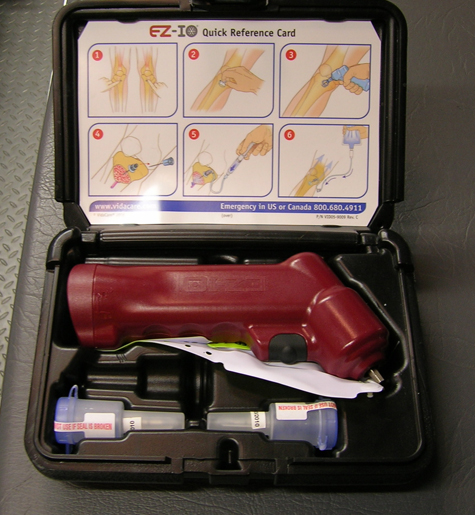
The second table shows IO flow rates in sick human patients in Singapore. You can see the flow rates with a pressure bag compare to a 20 gauge peripheral iv infusion rate, and also compare favorably with CVC infusion rates. In many of these patients, they had both a tibial and humerus IO placed for resuscitation.
Some pearls regarding IO access:
NO!
Until next time, I’ll just be standing on the corner, minding my own business.


Leave a Reply